Surgeons at The Prince Charles Hospital in Brisbane, Australia, recently carried out an incredibly complex procedure after discovering that a patient’s aorta had expanded to about four times its normal size.ABC News Australia was the first to report that the patient, a man in his late 50s, was living with what medical staff called a “ticking time bomb” in his chest.To prepare for the surgery, doctors used a life-sized 3D printed model of the man’s aorta.
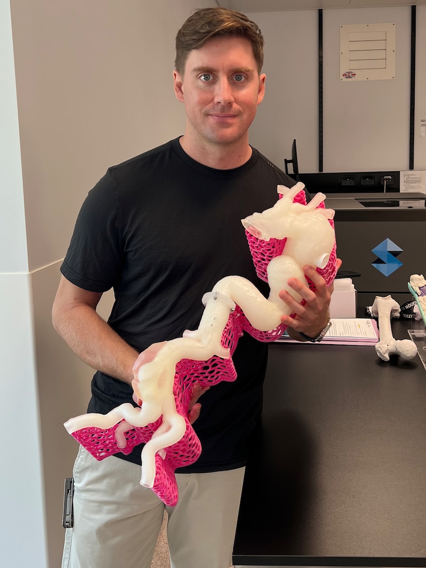
Created by engineers at the Herston Biofabrication Institute, part of Brisbane’s public hospital network, the model captures both the shape and flexibility of the artery.It took nearly four days to print using multiple materials so that surgeons could feel and study it before the operation.Liam Georgeson holds the life-size model used by surgeons to better plan the life-saving surgery.
Image courtesy of Queensland Health.There are 39 hospital units across Australia and New Zealand capable of performing adult cardiac surgery, but only a small number of these carry out highly complex procedures, such as full aortic arch replacements, especially using 3D printed planning models.The procedure, known as a total aortic arch replacement, is far from routine; it lasts around nine hours and requires the patient’s body to be cooled while blood flow is stopped for about 20 minutes.
During that time, surgeons work quickly to install a synthetic graft, which Dr Samantha Peden, a vascular and endovascular surgeon at The Prince Charles Hospital, compared to a “flexible, waterproof jacket.” Dr.Peden, who led the operation, called it “fancy plumbing.” According to the hospital, the patient is recovering well.Nearly three weeks later, he remained in the hospital but had left the intensive care unit and begun rehabilitation.
Reports included that there were no signs of paraplegia (a condition that causes paralysis of the lower half of the body), and the patient could use his arms and legs normally.He still needs another surgery to replace the lower part of his aorta later this year.This graft was used to replace most of a patient’s aorta in surgery in June.
Image courtesy of Queensland Health.Traditionally, surgeons rely on 2D scans, like CT or MRI images, for planning.But with a true-to-life 3D model, they can hold, rotate, and examine the exact shape of the patient’s artery.
This hands-on approach helps them plan more precisely for tricky curves, fragile walls, and blood vessel placements.According to industrial designer Liam Georgeson at Herston Biofabrication Institute, the model can combine hard and soft materials.That means some parts feel rigid, like bone, and others feel soft, like flesh, helping surgeons rehearse an operation.
The 3D printed aorta used in this case was produced on a Stratasys J750 Digital Anatomy Printer, a machine supplied by Objective3D, one of Australia’s leading providers of advanced 3D printing technology.In a social media post, Objective3D highlighted the surgery as a strong example of how realistic anatomical models can improve patient outcomes by giving surgeons the chance to rehearse complex procedures in advance.The Herston Biofabrication Institute, a medical customer of Objective3D, created the model and continues to collaborate with The Prince Charles Hospital to expand the use of this technology in surgical planning.
Dr Peden said, “It’s a real‑world example of health innovation translating directly into improved clinical practice.” Soft visual anatomy 3D printed models.Image courtesy of Stratasys.This isn’t the hospital’s first experience with 3D printing for heart surgery.
According to a research report by the hospital foundation, their Cardiothoracic Surgery Research Unit has been a leader in 3D printed aortic devices for years.In 2022, they supported over 100 patients with external 3D printed aortic root support devices under the PEARS (Personal External Aortic Root Support) program.PEARS uses personalized 3D printed meshes to reinforce weak parts of the natural aorta, and has improved outcomes for patients with aortic insufficiency or aneurysm risk.
The report also mentions the introduction of an Ascyrus Medical Dissection Stent and collaboration across major universities, showing a commitment to innovation in aortic care.Inside an operating theatre at The Prince Charles Hospital, where surgeons worked to replace most of a man’s aorta.Image courtesy of Queensland Health.
This case is important because it demonstrates how advanced 3D printing can improve patient safety.By giving surgeons a physical model of the patient’s aorta ahead of time, they were better prepared for what they would face in the operating room (OR), reducing surprises and guiding surgical decisions.In fact, the patient’s smooth recovery, without major complications like paralysis, is even more proof of the importance of this type of pre-planning procedure.
It shows how tools like 3D printed replicas can help lead to better outcomes, even in the most complex and high-risk procedures.Although everything worked out ideally, the Herston Biofabrication Institute is continuing to develop even more advanced models that include simulated blood flow, letting doctors practice on lifelike, pulsating replicas before actual surgeries.Subscribe to Our Email Newsletter Stay up-to-date on all the latest news from the 3D printing industry and receive information and offers from third party vendors.
Print Services
Upload your 3D Models and get them printed quickly and efficiently.Powered by FacFox
Powered by 3D Systems
Powered by Craftcloud
Powered by Endeavor 3D
Powered by Xometry
3DPrinting Business Directory
3DPrinting Business Directory